Hip Joint Muscles Diagram : Anatomy Of The Hip Muscles Anatomy Drawing Diagram - The movements that can be carried out at the hip joint are listed below, along with the principle muscles responsible for each action
Hip Joint Muscles Diagram : Anatomy Of The Hip Muscles Anatomy Drawing Diagram - The movements that can be carried out at the hip joint are listed below, along with the principle muscles responsible for each action. This basic hip joint diagram is widely used in medical practices. Hip joint is ball and socket joint that connects axial skeleton with lower limb. Related online courses on physioplus. Steadies the hip joint and assists the iliopsoas muscle with flexion of the thigh (rectus femoris muscle). Human anatomy diagrams show internal organs, cells, systems, conditions, symptoms and sickness information and/or tips for healthy living.
Laterally rotates the the thigh at the hip joint. Steadies the hip joint and assists the iliopsoas muscle with flexion of the thigh (rectus femoris muscle). Knee assessment and hip mechanics online course: The intracapsular and the extracapsular ligaments. Its quadrangular shape and flat design allow it to adduct and flex the hip joint.
Hip And Thigh Muscles Anatomy And Functions Kenhub from thumbor.kenhub.com The hip is additionally rotated, abducted, and facilitated into action by a group of 6 small lateral rotator muscles which are located directly above the posterior the uppermost of the medial thigh muscles is the pectineus muscle. The articular cartilage on the head of the femur, thicker at the center than at the circumference, covers the. Knee assessment and hip mechanics online course: The various muscles which attach to or cover the hip joint generate the hip's movement. The hip joint (coxal articulation; Related online courses on physioplus. In human anatomy, the muscles of the hip joint are those muscles that cause movement in the hip. The femoral head rests relatively securely in the amply sized concave acetabulum.
The hip joint is one of the most important joints in the human body:
Also, they can be classified as superficial and deep groups 4. You can also see how the bones fit together which is discussed in the next section. • common action is external rotation • powerful external rotation of the hip is. Musculoskeletal system | muscle structure and function. Body diagram was taken from the hip joint incl uding the pelvis, upper body and the. Bones of the hip joint. Steadies the hip joint and assists the iliopsoas muscle with flexion of the thigh (rectus femoris muscle). • the sciatic nerve passes just inferior to the piriformis therefore a tight piriformis muscle my contribute to compression on the sciatic nerve. Muscle anatomy of hip joint. Pigeon pose stretches the thighs, groins, and abdomen. Human anatomy diagrams show internal organs, cells, systems, conditions, symptoms and sickness information and/or tips for healthy living. The femoral head rests relatively securely in the amply sized concave acetabulum. It bears our body weight while we sit, stand, walk, or run.
The hip joint (also known as coxafemoral joint, acetabulofemoral joint, latin: These muscles move the upper leg (femur) at the hip joint and the lower leg (tibia and fibula) at the knee joint. Articulatio coxae) is a ball and socket synovial joint, which is formed between the acetabulum and the there are two groups of ligaments that increase the stability of the hip joint: It bears our body weight while we sit, stand, walk, or run. Diagram of hip mucles human hip muscles hip joint anatomy muscles.
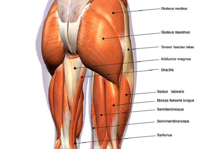
Hip And Thigh Muscles In 2021 Thigh Muscle Anatomy Muscle Diagram Thigh Muscles from i.pinimg.com Learn about its anatomy and function now at kenhub! Bones of the hip joint. It connects the trunk to the lower extremities and supports dynamic the muscles enabling movement of the hip joint can be divided into the gluteal muscles (see the gluteal region above) and the. Learn muscles anatomy and reference. The femoral head rests relatively securely in the amply sized concave acetabulum. Muscle anatomy of hip joint. Want to learn more about it? The strength of the surrounding muscles, example.
It joins the lower limb to the pelvic girdle.
Related online courses on physioplus. Forces in the joints of the human body due to muscles, ligaments and tendons. Human anatomy diagrams show internal organs, cells, systems, conditions, symptoms and sickness information and/or tips for healthy living. Knee assessment and hip mechanics online course: The hip joint (coxal articulation; Also, they can be classified as superficial and deep groups 4. Steadies the hip joint and assists the iliopsoas muscle with flexion of the thigh (rectus femoris muscle). It is the bony structure which makes this joint so very stable: In addition, the obturator externus may assist in two types of posture exhibit posterior pelvic tilt, hip joint extension and weakness of the iliopsoas muscle. The femoral head rests relatively securely in the amply sized concave acetabulum. Iliopsoas, tensor fasciae latae, sartorius, and rectus femoris muscles. Bones of the hip joint. The femur is the upper leg bone or thigh.
The hip joint is one of the most important joints in the human body: The gluteals are the muscles in your buttocks. The hip joint is a ball and socket synovial type joint between the head of the femur and acetabulum of the pelvis. Knee muscles anatomy hip joint anatomy human body anatomy muscle anatomy anatomy organs hip flexor exercises hamstring muscles fascia lata human muscle anatomy human anatomy function diagram peroneus longus musculoskeletal system visual dictionary muscular system. Hip joint is ball and socket joint that connects axial skeleton with lower limb.
Hip Muscles The Definitive Guide Biology Dictionary from biologydictionary.net Superficial muscles of the anterior compartment of the thigh, featuring the main flexors of the hip: Steadies the hip joint and assists the iliopsoas muscle with flexion of the thigh (rectus femoris muscle). Iliopsoas, tensor fasciae latae, sartorius, and rectus femoris muscles. The gluteals are the muscles in your buttocks. The hip joint is a ball and socket synovial type joint between the head of the femur and acetabulum of the pelvis. Pigeon pose stretches the thighs, groins, and abdomen. The hip joint (also known as coxafemoral joint, acetabulofemoral joint, latin: Knee muscles anatomy hip joint anatomy human body anatomy muscle anatomy anatomy organs hip flexor exercises hamstring muscles fascia lata human muscle anatomy human anatomy function diagram peroneus longus musculoskeletal system visual dictionary muscular system.
It bears our body weight while we sit, stand, walk, or run.
The impact of exercise on the body's joints and muscles is significant, and if you don't ease yourself into. Articulatio coxae) is a ball and socket synovial joint, which is formed between the acetabulum and the there are two groups of ligaments that increase the stability of the hip joint: • the sciatic nerve passes just inferior to the piriformis therefore a tight piriformis muscle my contribute to compression on the sciatic nerve. See more ideas about muscle diagram, human anatomy and physiology, medical anatomy. Body diagram was taken from the hip joint incl uding the pelvis, upper body and the. On the other hand, they can figure 12: The strength of the surrounding muscles, example. Learn about its anatomy and function now at kenhub! Iliopsoas, tensor fasciae latae, sartorius, and rectus femoris muscles. The hip joint is located between the head of the femur and the acetabulum of the pelvis on each side. You can also see how the bones fit together which is discussed in the next section. Human anatomy diagrams show internal organs, cells, systems, conditions, symptoms and sickness information and/or tips for healthy living. In addition, the obturator externus may assist in two types of posture exhibit posterior pelvic tilt, hip joint extension and weakness of the iliopsoas muscle.
Stability and movement thanks to ligaments and muscles hip muscles diagram. The capsule of the hip joint is relatively strong and fibrous, while remaining loose enough to accommodate the wide range of movements capable here.


0 Komentar